In a significant breakthrough, a research team led by Professor Fan Yubo, Professor Du Jing, and Associate Professor Yao Jie from the School of Biological Science and Medical Engineering at Beihang University has uncovered the mechanisms driving embryonic pattern formation during early mammalian embryonic development.
Their findings, published in the prestigious journal Cell Reports under the title of “Cavity oscillation drives pattern formation in early mammalian embryos,” address a long-standing scientific mystery: how the inner cell mass (ICM) separates into the spatially distinct epiblast (EPI) and primitive endoderm (PrE) during the second cell fate in mammalian embryogenesis.
Professors Fan Yubo and Du Jing serve as the co-corresponding authors of the study. The co-first authors include Guo Zheng, a Ph.D. candidate at Beihang University, Associate Professor Yao Jie, and Associate Researcher Zheng Xu from the Institute of Mechanics, Chinese Academy of Sciences. The study also benefited from the significant contributions of Professor Li Lei from the Institute of Zoology, Chinese Academy of Sciences, Professor Feng Yue from Beijing University of Chemical Technology, and Researcher Guan Dongshi and Associate Researcher Li Long from the Institute of Mechanics, Chinese Academy of Sciences. Beihang University is recognized as the primary affiliation for this research.
Using a combination of innovative experimental techniques and computational simulations, the team investigated the role of blastocyst cavity oscillation in embryo patterning. They discovered that during the mid-blastocyst development, periodic oscillations of the blastocyst cavity are essential for the movement of pPrEs toward the ICM-lumen interface (Figure 1).
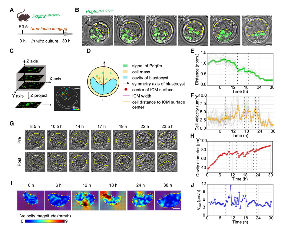
Figure 1. Movement of pPrEs toward the ICM-lumen interface accompanied by strong cyclic embryo oscillation in mid-blastocyst
By manipulating the oscillation and expansion of the blastocyst cavity through biological and mechanical methods, the researchers found that inhibiting cavity oscillation delayed the spatial separation of pEPIs and pPrEs (Figure 2), while enhanced cavity oscillation accelerated the process (Figure 3).
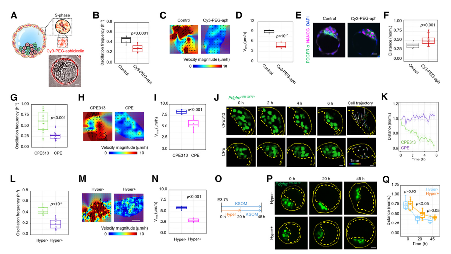
Figure 2. The spatial segregation of pEPIs and pPrEs requires strong cyclic oscillation of the mid-blastocyst cavity
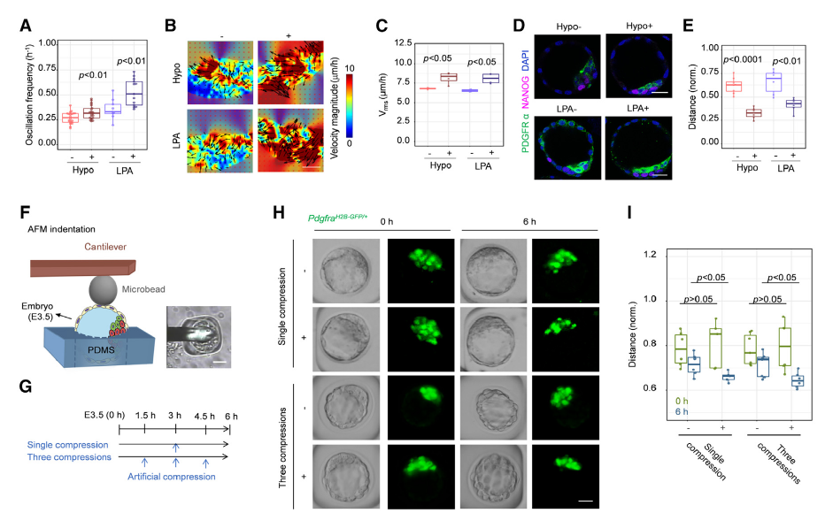
Figure 3. Enhanced cavity oscillation facilitates the spatial segregation of pEPIs and pPrEs in mid-blastocyst
The study elucidated the mechanisms underlying this phenomenon, showing that reducing the difference in the surface properties (i.e., surface tension fluctuation and affinity) between the two types of simulated cells attenuated the segregation process. Numerical simulations and experimental validation confirmed the essential role of cavity oscillation in promoting cell motility and segregation (Figure 4).

Figure 4. Blastocyst cavity oscillation drives EPI/PrE segregation by promoting cell-cell contact fluctuations of ICM cells
The research team further explored the PrE epithelialization in late blastocyst. Their findings showed that the transition is primarily driven by the sustained expansion of the late blastocyst, while cavity oscillation, a significant factor in earlier stages, plays a reduced role in this phase (Figure 5).
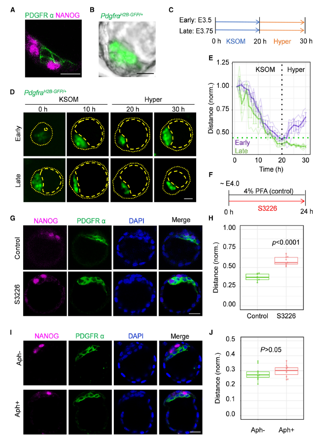
Figure 5. Cavity expansion after EPI/PrE segregation is required for PrE epithelialization in late blastocyst
The study also examined the impact of blastocyst cavity mechanics on gene expression in ICM cells. Experimental results demonstrated that cavity pressure and cavity oscillation jointly enhance DGFRα expression and YAP nuclear accumulation in pPrEs, highlighting their critical role in cell fate determination (Figure 6).
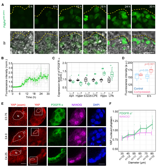
Figure 6. PDGFRα expression and YAP nuclear accumulation in pPrEs are increased in response to blastocyst cavity pressure and oscillation
The research not only deepens our understanding of the mechanisms behind embryo pattern formation but also pioneers new pathways for exploring the role of physical forces in developmental biology. Its findings have profound implications for guiding the design and construction of artificial tissues and accelerating advancements in clinical applications.
This work was supported by the National Natural Science Foundation of China, the National Key R&D Program of China, Young Elite Scientist Sponsorship Program by CAST , the CAS Strategic Priority Research Program, Fundamental Research Funds for the Central Universities, and the Youth Innovation Promotion Association CAS.
Link to the article: https://doi.org/10.1016/j.celrep.2025.115342
Editor: Lyu Xingyun