Professor Zhao Wei's team from the School of Physics at Beihang University has made significant strides in low-dose computed tomography (LDCT) imaging. Their latest study, titled "NIFA: Low-dose CT imaging via noise intensity field aware networks," has been published in the prestigious international journal Medical Image Analysis. This work introduces a novel denoising network, NIFA, which effectively reduces radiation dose while preserving the diagnostic value of computed tomography (CT) images.
CT is one of the most widely used imaging modalities in clinical practice. While highly effective for disease diagnosis, the extensive use of CT has contributed to the major part of population-based radiation dose, raising public concerns about the potential risk of cancer. Therefore, LDCT has attracted much attention in the past decades. LDCT lowers x-ray dose to reduce health risks during data acquisition, but it introduces excessive image noise and artifacts, compromising image quality, which hinders its clinical use.
To address this problem, the researchers investigate data-driven deep neural networks to improve LDCT image quality. They tackle the LDCT challenge by leveraging the data-driven paradigm and CT imaging physics to develop a more clinically relevant predictive model. They formulate noise in LDCT images as a noise intensity field and denoising process as intensity value regression. Based on the formulation, a noise intensity field aware (NIFA) network which separately extracts low-intensity and high-intensity information is proposed to reduce the magnitude of the intensity field while preserving texture information and anatomical details.
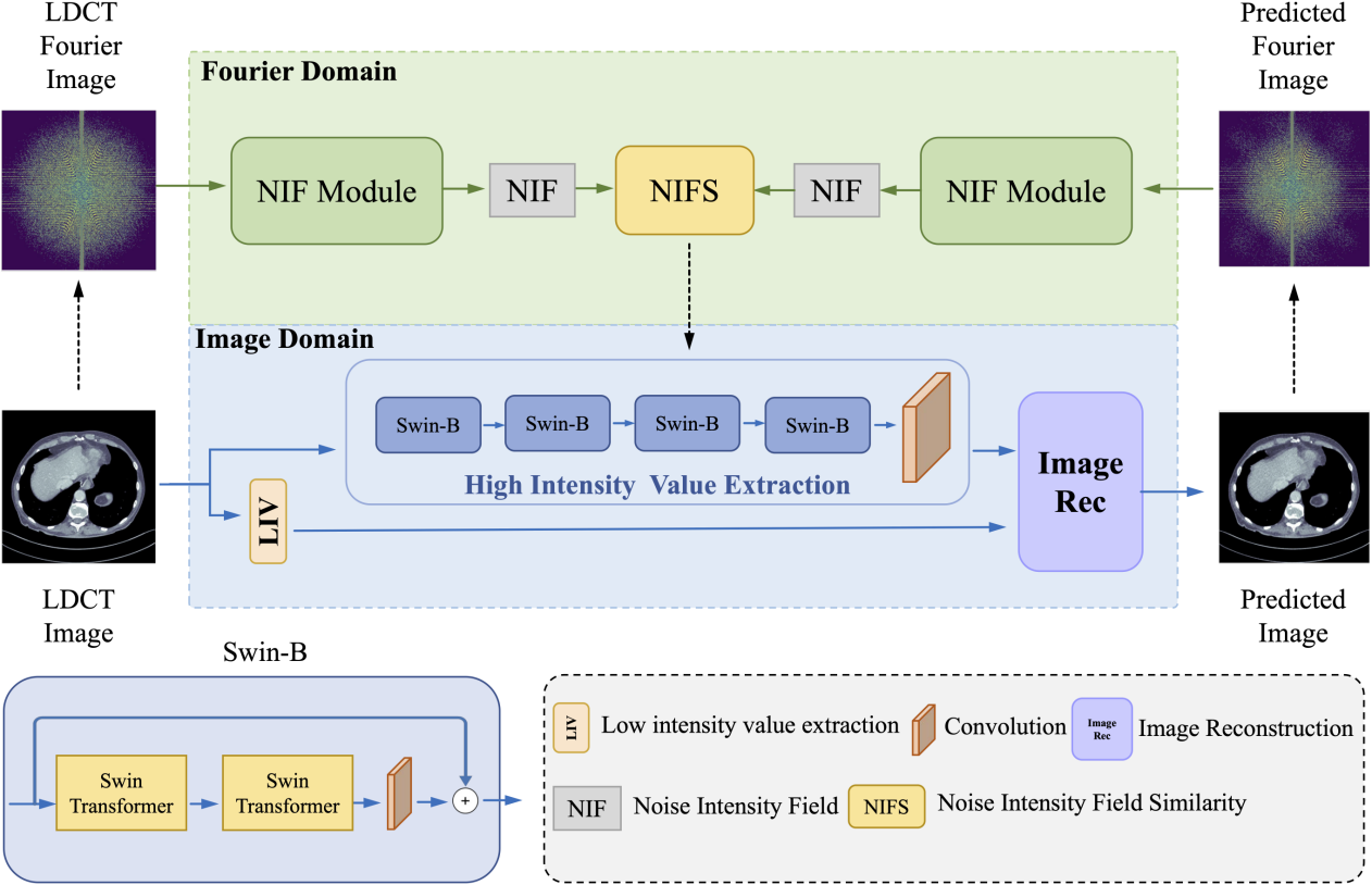
Fig.1. The pipeline of the NIFA network
In the study, extensive experiments were conducted on both the AAPM dataset and numerical simulated dataset constructed by the team to validate the performance of the proposed method. Ablation studies were also performed to demonstrate the effectiveness of the design. Clinically relevant metrics, including the noise power spectrum, were used to quantitatively evaluate the quality of the CT images.
Beyond low-dose medical CT imaging, the NIFA network offers promising applications in industrial non-destructive testing scenarios that require rapid scanning coupled with high texture fidelity, indicating broad engineering potential.
The paper's co-first authors are Zhao Zihui (a 2023 graduate of Beihang University, now a Master's student at Tsinghua University) and Wang Yanxin (a Ph.D. student at the Hangzhou International Innovation Institute of Beihang University). Professor Zhao Wei from Beihang University and Professor Li Xiaomeng from the Hong Kong University of Science and Technology are the corresponding authors. Beihang University is the primary affiliation for the study. Tian Suqing, Associate Chief Physician from Peking University Third Hospital, also contributed to this research.
The study received support from the National Natural Science Foundation of China, the Natural Science Foundation of Zhejiang Province, and the Fundamental Research Funds for the Central Universities, China.
Medical Image Analysis, established in 1996 and published by Elsevier, is a leading international journal in the field of medical and biological image analysis. With a recent three-year average impact factor of 11.8, it holds significant academic influence within the medical imaging community.
Link to the article: https://doi.org/10.1016/j.media.2025.103866
Editor: Lyu Xingyun


 Beihang News8th China Microelectronics Intelligent Conference convenes in BeijingMore
 Beihang NewsBeihang AI Knowledge Center debuts in MexicoMore
 Beihang News18th Enterprise Open Day successfully heldMore
 Beihang NewsProfessor Niu Jianwei elected as IEEE FellowMore
 Beihang NewsTen Beihang professors and alumni elected as CAS and CAE academiciansMore
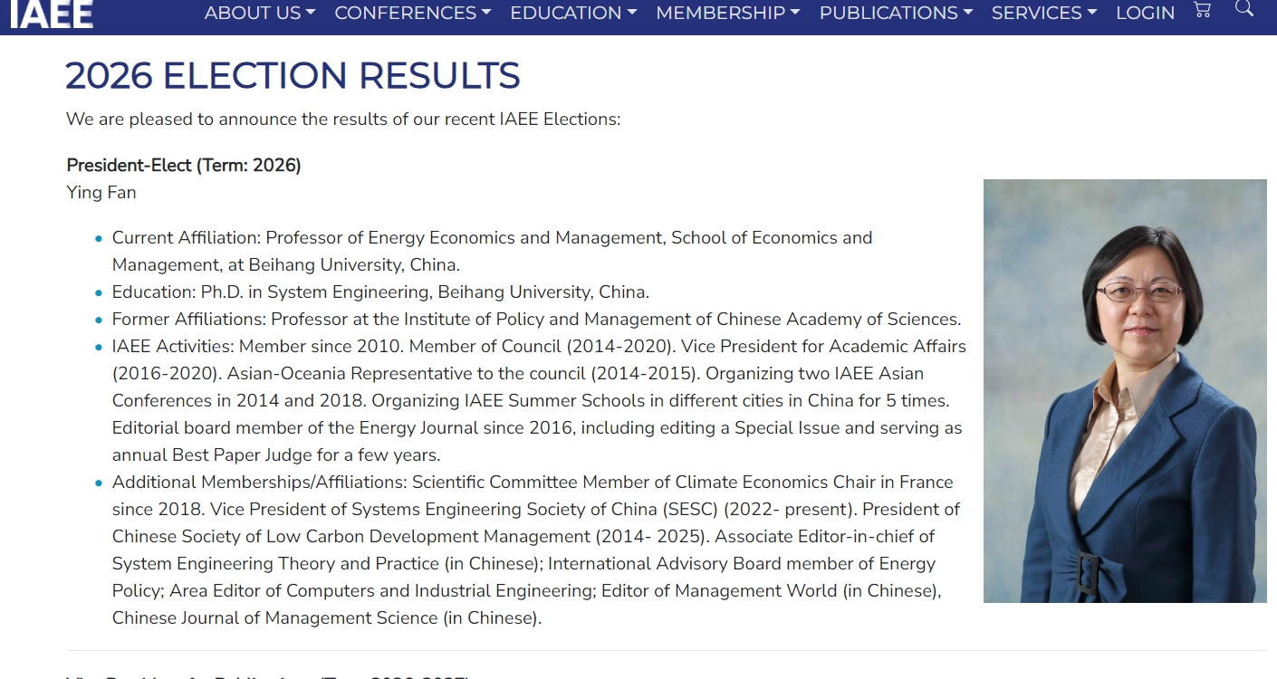 Beihang NewsProfessor Fan Ying named IAEE President-ElectMore